👉 Learn how to build AI systems for medical imaging domain by leveraging tools and techniques that I share with you! | 💡 The newsletter is read by people from: Nvidia, Baker Hughes, Harvard, NYU, Columbia University, University of Toronto and more!
Visualize Medical Data in the Browser?
Hi Reader,,
Welcome to the PYCAD newsletter, where every week you receive doses of machine learning and computer vision techniques and tools to help you learn how to build AI solutions to empower the most vulnerable members of our society, patients.
Visualize 3D Medical Data in the Browser using Python
One of our latest experiments at PYCAD was to research how we can visualize 3D data on the browser by directly using Python.
This can help us build web apps for POCs in medical imaging.
One great tool that we recently discovered is called stpvista.
This is in fact a tool that allows you to integrate Pyvista plots, which is a great Python library for 3D visualization, directly inside your Streamlit web application.
Below you can see an example of visualizing an STL file of an intraoral scan on the browser using Python and Streamlit!
Pyvista is already a huge library for 3D visualization, so having the possibility to integrate its plots inside a Streamlit web app is just the cherry on top of the cake!
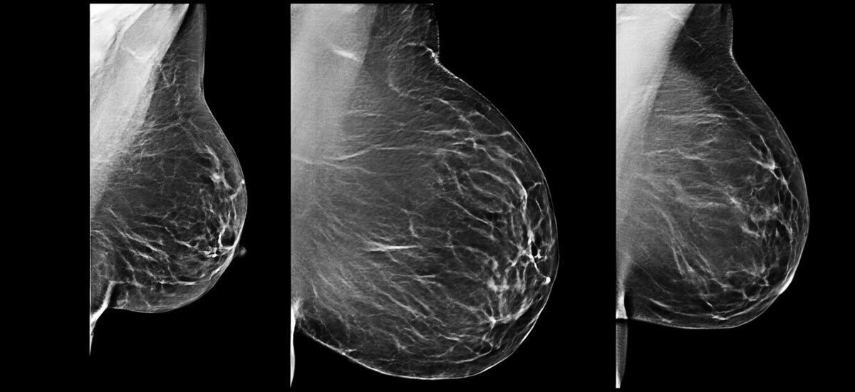
Medical Imaging Data Formats
I work daily on medical imaging problems. Here are 3 file formats that we deal with constantly.
Let’s demystify these 3 common formats: DICOM, NIFTI, and NRRD.
DICOM (Digital Imaging and Communications in Medicine)
- Industry Standard: Used primarily in radiology and often by imaging devices directly.
- Rich Metadata: Contains patient info, imaging parameters, and acquisition details.
- Structure: Packs multiple 2D slices to form a 3D image.
- Usage: Ideal for clinical applications and PACS integration.
NIFTI (Neuroimaging Informatics Technology Initiative)
- Brain Imaging: Primarily designed for the neuroimaging community.
- Structure: Single file storage of 3D or 4D data (includes time-series data).
- Header Info: Contains spatial and temporal metadata.
- Usage: Research-focused, especially functional MRI studies.
NRRD (Nearly Raw Raster Data)
- Flexibility: Supports a broad range of medical image data types.
- Header-Body Separation: Can keep metadata separate from image data.
- Extensibility: Open format with easy-to-adapt properties.
- Usage: Advanced visualization applications and research purposes.
In summary, the choice of format often depends on the application. DICOM is great for clinical setups, NIFTI shines in research, especially neuroimaging, and NRRD offers flexibility for diverse visualization needs.
Stay informed, and choose wisely based on your specific requirements!
We Can Help You with Your Next Medical Imaging ProjectIf your company or organization is looking to build a machine learning solution for a medical imaging problem, then feel free to reach out to us at: We can help you build a full ML solution from training to deployment with affordable rates! You can check out some of the projects that we worked on here: https://pycad.co/portfolio and some of our clients case studies here. |
Tweet of the Day
|

That's it for this week's edition, I hope you enjoyed it!
Machine Learning for Medical Imaging
by Nour Islam Mokhtari from pycad.co
👉 Learn how to build AI systems for medical imaging domain by leveraging tools and techniques that I share with you! | 💡 The newsletter is read by people from: Nvidia, Baker Hughes, Harvard, NYU, Columbia University, University of Toronto and more!
Hi Reader,, Welcome to the PYCAD newsletter, where every week you receive doses of machine learning and computer vision techniques and tools to help you learn how to build AI solutions to empower the most vulnerable members of our society, patients. TotalSegmentator : Whole Body Segmentation at your Fingertips This free tool available online can do full body segmentation, it's called TotalSegmentator. I have already mentioned this tool in a previous edition of the newsletter, but in this...
Hello Reader, Welcome to another edition of PYCAD newsletter where we cover interesting topics in Machine Learning and Computer Vision applied to Medical Imaging. The goal of this newsletter is to help you stay up-to-date and learn important concepts in this amazing field! I've got some cool insights for you below ↓ A Medical Imaging Expert Told Me This Recently I saw a post on LinkedIn where a medical imaging expert showcased his work of segmenting the lungs and its bronchial trees. You can...
Hello Reader, Welcome to another edition of PYCAD newsletter where we cover interesting topics in Machine Learning and Computer Vision applied to Medical Imaging. The goal of this newsletter is to help you stay up-to-date and learn important concepts in this amazing field! I've got some cool insights for you below ↓ How we helped accelerate inference time for a client's AI product Below is a screenshot of a benchmark we did for a client of ours. The goal was to accelerate inference time. This...