👉 Learn how to build AI systems for medical imaging domain by leveraging tools and techniques that I share with you! | 💡 The newsletter is read by people from: Nvidia, Baker Hughes, Harvard, NYU, Columbia University, University of Toronto and more!
ML for Surgical Planning
|
Hello Reader, Welcome to another edition of PYCAD newsletter where we cover interesting topics in Machine Learning and Computer Vision applied to Medical Imaging. The goal of this newsletter is to help you stay up-to-date and learn important concepts in this amazing field! I've got some cool insights for you below ↓ |
Case study: ML for Surgical Planning
Did you know that before you have a surgery, your medical team will do what is called a surgical plan?
What is it and how can machine learning help with this?
Surgical planning is a preparatory process undertaken by medical professionals, especially surgeons, before a surgical procedure.
It involves a series of steps designed to gather information about the patient's condition and anatomy, develop a detailed strategy for the operation, anticipate potential complications, and devise contingency plans to deal with them.
Key elements of surgical planning often include:
1 - Reviewing the patient's medical history and current health status.
2 - Analyzing diagnostic tests and imaging studies (like CT scans, MRIs, or X-rays) to understand the specifics of the patient's condition and the surgical site.
3 - Creating a 3D model or visual representation of the area to be operated on, often by using computer software.
4 - Segmenting the area of interest in the 3D model, identifying key structures or anomalies.
5 - Deciding on the best surgical approach and techniques to use, based on all the gathered information.
6 - Anticipating potential complications and formulating strategies to mitigate them.
The goal of surgical planning is to increase the predictability, safety, and efficiency of the operation, ultimately leading to improved patient outcomes.
In most medical clinics and hospitals, the 4th step is still being done manually!
This means that a medical practitioner or technician has to go through MRI or CT scans slice by slice. In each slice he or she has to manually highlight the region of interest.
If you ever worked with 3D medical data before, then you'd know that this is a tedious task.
An MRI can have hundreds of slices.
This is where deep learning can play an important role. Imagine if a medical professional can automatically segment hundreds of patient medical data. And for each patient, hundreds of MRI slices can be automatically segmented! This is a clear win for any medical professional!
Why do you need to know this?
Several medical practitioners are unaware that AI and machine learning can help them boost the performance of their business or practice. In surgical planning it's currently the case.
Several medical practitioners are still doing a lot of the annotation of their 3D data manually. So if you're working in the field of medical imaging, then maybe you can share the insights I mentioned above to your colleagues or to your IT team. If you can't convince them yourself, then feel free to reach out to me. I'll be glad to offer a free one hour consulting to show your medical team why they should definitely start implementing AI!
Big Tech Companies and Healthcare
Big tech is going strong on machine learning for the healthcare industry. Here’s one tool from Microsoft that I haven’t seen people talk about yet.
The tool is a python package called HI-ML.
HI-ML is a deep learning toolbox for medical imaging. It has a seamless integration with Azure.
This toolbox contains several functionalities that alleviates some of the difficulties of using deep learning for medical imaging.
Examples:
- You can do self-supervised learning to do pre-training of an image encoder for medical images.
- You can run training on a local GPU machine or inside of AzureML without code changes.
- It allows working with whole slide images for medical pathologies.
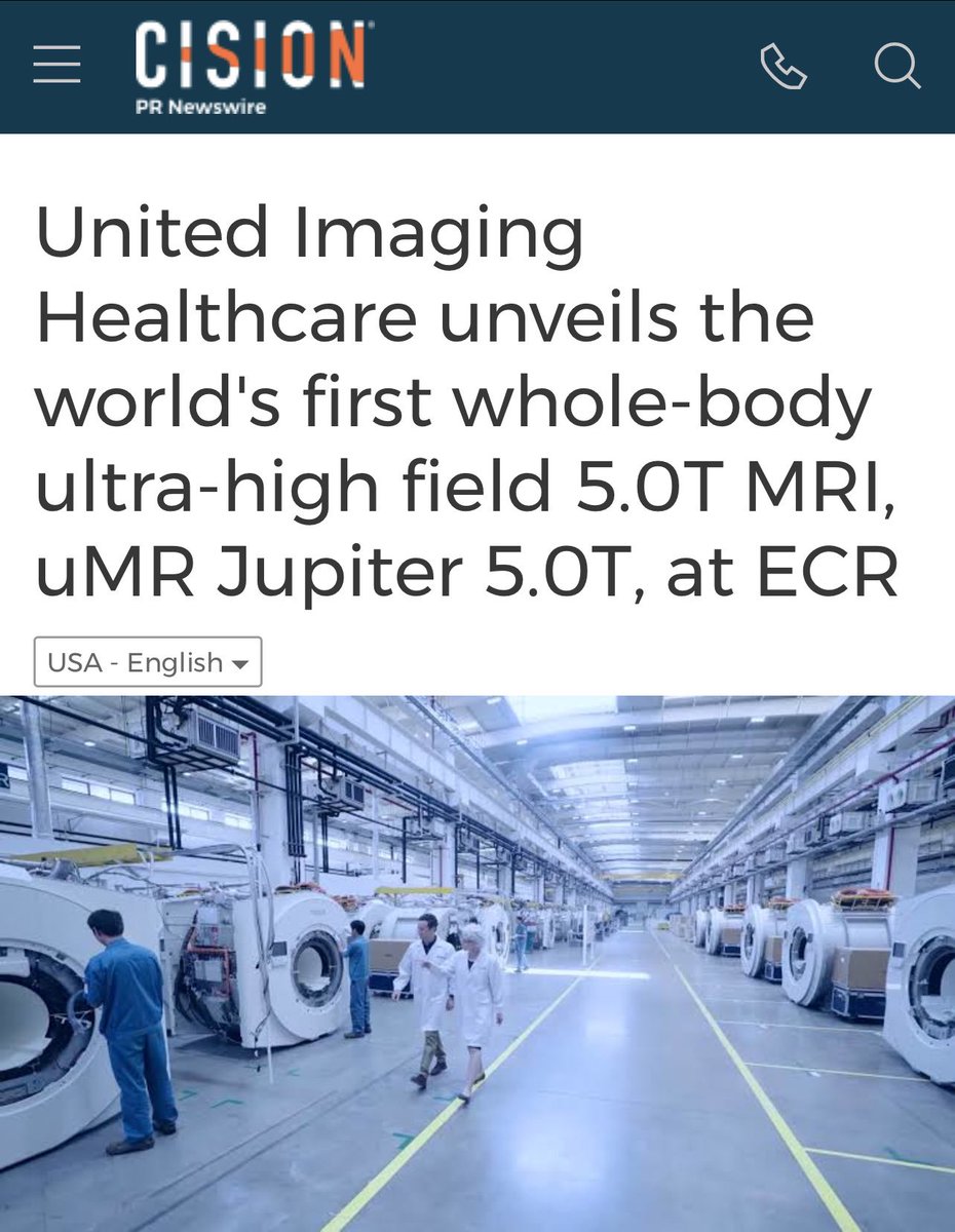
China is now leading innovation when it comes to medical imaging devices
|

That's it for this week's edition, I hope you enjoyed it!
Machine Learning for Medical Imaging
👉 Learn how to build AI systems for medical imaging domain by leveraging tools and techniques that I share with you! | 💡 The newsletter is read by people from: Nvidia, Baker Hughes, Harvard, NYU, Columbia University, University of Toronto and more!
Hello Reader, Welcome to another edition of PYCAD newsletter where we cover interesting topics in Machine Learning and Computer Vision applied to Medical Imaging. The goal of this newsletter is to help you stay up-to-date and learn important concepts in this amazing field! I've got some cool insights for you below ↓ AI Scribes: Transforming Medical Documentation Web Application for Medical Note Generation AI-powered medical scribes are revolutionizing clinical workflows by automating...

Hello Reader, Welcome to another edition of PYCAD newsletter where we cover interesting topics in Machine Learning and Computer Vision applied to Medical Imaging. The goal of this newsletter is to help you stay up-to-date and learn important concepts in this amazing field! I've got some cool insights for you below ↓ DeepSeek: A New Player in AI for Healthcare The new open-source LLM, DeepSeek, is creating buzz for its potential to transform AI in medicine and healthcare. Designed for...
Hello Reader, Welcome to another edition of PYCAD newsletter where we cover interesting topics in Machine Learning and Computer Vision applied to Medical Imaging. The goal of this newsletter is to help you stay up-to-date and learn important concepts in this amazing field! I've got some cool insights for you below ↓ Now You Can Use Large Language Models that are HIPAA Compliant People are finding ways to use large language models in all fields. MedTech is no exception. The amount of work...